Multi-modal
Built for X-ray, CT, MRI, ultrasound, fundus, dermoscopy, histology and angiography, with a specialist agent for each imaging domain.
Intelligent model selection, automated segmentation and independent quality verification across medical imaging domains.
Start with $10 of free credit. No card needed.
Built for X-ray, CT, MRI, ultrasound, fundus, dermoscopy, histology and angiography, with a specialist agent for each imaging domain.
The right model for every task: specialised segmentation models and foundation models, chosen by the agents, with a library that keeps growing.
An independent reviewer checks every mask. Uncertain or low-confidence results are flagged for expert review instead of slipping through.
How it works
A team of AI agents does the work an annotation team would: read the image, pick the tools, draw, check, and send anything uncertain to a person.
Upload your images and say what to annotate, in plain words.
An agent identifies the modality, the anatomy and the annotation task.
The orchestration layer picks the most suitable segmentation model for the task.
Models draw the masks; agents refine them piece by piece.
A separate reviewer checks each mask, names its errors and sends it back to be fixed.
Uncertain cases go to human experts; accepted results are ready to export.
A real run
A public chest X-ray and one request: Outline the pneumothorax
. The
reviewer rejected the first two masks and said why; the third was accepted. Nothing here is
retouched.
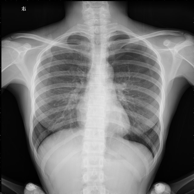
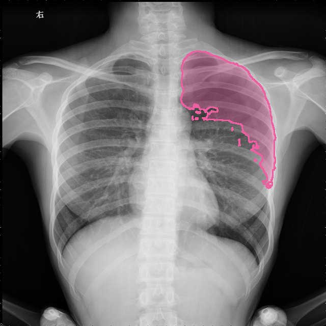
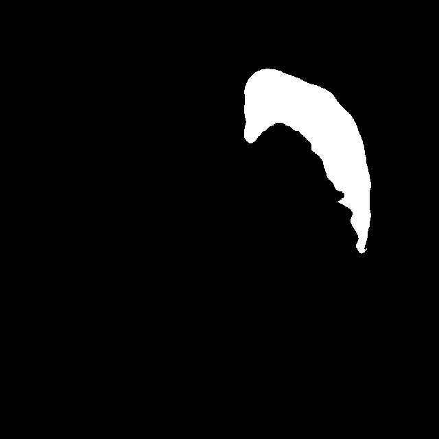
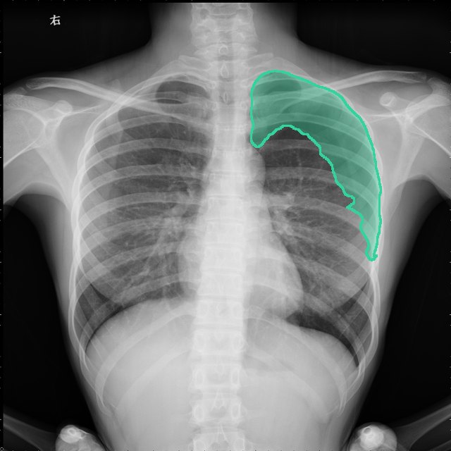
The lung-side boundary extends below the visible visceral pleural line into vascularized left upper lung.
Structure 1 includes a substantial band of vascularized upper-left lung.
The mask covers the left apical and lateral pneumothorax, broadly following the visceral pleural boundary and inner chest wall.
Image and expert examples: PTX-498 (Wang et al.), CC BY 4.0; masks drawn by trustmed AI. Scores are the reviewer's own, from 0 to 1.
Who it's for
Label training data faster, with every mask checked before it reaches your model.
Turn image collections into annotated datasets without months of manual outlining.
Annotate across modalities for studies and theses, with the reasoning behind every mask.
Start a new task from a handful of expert examples and grow specialised models from it.
Every step recorded and reviewable, with experts in the loop where it matters.
Privacy and security
Contact
A pilot on your data, a demo, a research collaboration, or anything else: the team answers by email. Or write to [email protected].